| Main Page |
|
Capacitance |
|
Polarization | Cursos |
|
Neil C. Bruce | |||||
|
Monte Carlo
calculations of scattering in the human forearm
In the study of the scattering of light in tissues for medical applications, it is almost always assumed that the tissue is homogeneous. However, many tissue materials are fibrous. The most common examples of this are muscle and brain tissue. In this type of tissue the scattering properties of the medium depend on the direction of propagation of the light. There may be two different scattering coefficients and two different absorption coefficients for the same tissue.
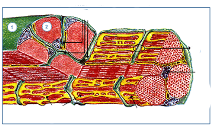
The above figure shows a schematic drawing of muscle tissue showing the different fibrous structures which make up this tissue. Muscle consists of packets of fibres where each fibre is between 20 and 100 mm in diameter, and the fibres are made up of cylindrical structures parallel to the fibres axes which are about 1 to 3 mm in diameter.
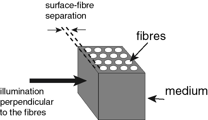
To show the effect of the fibres on the CW scattered light we modelled the situation shown in the figure below, for different distances between the fibres and the sample surface.
 |
|
|||||||||||||||||||||
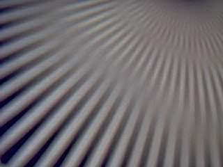
With this system we obtained the following simulation results, for the CW (continuous) light case.
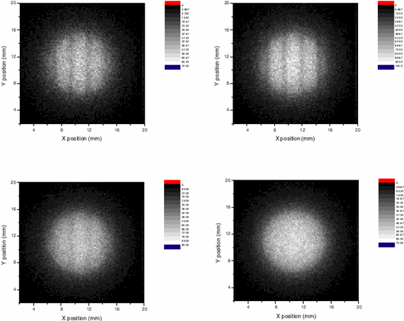
The graphs are for the cases of surface-fibre distances 0.04mm (top left); 0.08mm (top right), 0.4mm (bottom left) and 0.8mm (bottom right). Notice how the contrast in the shadow of the fibres disappears as the surface-fibre distance is increased.
To simulate scattering in fibrous tissue we modelled the human forearm as shown in the figure below.
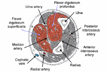

The figure on the left above is a schematic of the human forearm and on the right is the model used in the simulation. The parameters of the simulation are shown below.
|
|
Forearm
|
Radio
|
Ulna
|
|
Diameter (mm)
|
70.0
|
16.47
|
16.47
|
|
Position (mm)
|
0, 0
|
4.1, -10.3
|
12.3, 10.3
|
|
|
Muscle tissue
|
Muscle fibres
|
Fat Layer
|
Bone
|
|
Absorption Coefficient (1/mm)
|
0.03
|
0.03
|
0.005
|
0.027
|
|
Scattering Coefficient (1/mm)
|
16.0
|
11.4
|
20.0
|
36.0
|
|
g
|
0.95
|
0.95
|
0.95
|
0.95
|
|
n
|
1.4
|
1.4
|
1.4
|
1.4
|
|
Fibre radius (mm)
|
|
1.0
|
|
|
|
Fibre Separation (mm)
|
|
2.0
|
|
|
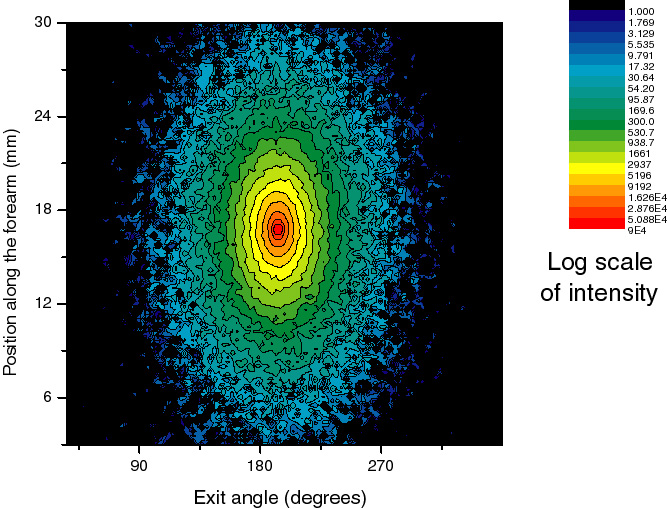
An example of the typical scattering pattern obtained in the model for CW light is shown below.
The equation used to fit to the numerical data is from the random-walk theory of Dagdug, Weiss y Gandjbakhche (Phys. Med. Biol., 48, 1361-1370, 2003) and is given by

where d is the width of the sample, ma and ms are the absorption and scattering coefficients and B is the anisotropy factor of the sample and depends on the difference in the scattering properties in two perpendicular directions. This is the new parameter for fibrous materials.
Results of the Monte Carlo program and the best fit of this equation are shown below. The points are the numerical results and the red lines are the theoretical results. The graphs are for in-plane reflection and for detection at different distances from the incident point: from 0.33mm (top left) to 0.51cm (bottom right). Note how the scattered pulses are longer for larger separations.
 |
 |
 |
 |
From the fit data, the following graphs were derived for the in-plane scattering for two different thicknesses of the fatty layer (0.355cm on the left and 0.01cm on the right) on the arm model and at two different scattering angles (180° corresponds to the straight through transmission direction). These graphs show the value of anisotropy parameter B.
 |
 |
From these graphs it can be seen that the effect of the anisotropy appears stronger when the separation between the incident point and the detector is larger, because the light travels further in the fibrous tissue and there is a larger effect on the scattered signal. It can also be seen that the thickness of the fatty layer and the detection position relative to the bones have very little effect on the parameter B.
The conclusion of this work is that the anisotropy is important for scattering in fibrous tissue, such as muscle.
This work was published in the Masters
thesis of Eglain Constantin Carrera (UNAM, 2007)
| ICAT |