| Main Page |
|
Capacitance |
|
Polarization | Cursos |
|
Neil C. Bruce | |||||
|
Scattering of light
in the human eye
Intraocular scattering has been known for some time to be an important problem. Even for healthy eyes the scattering from a bright source can mask the presence of accompanying objects. Scattering inside the eye can be increased with age, with disease, e.g. cataracts or by cellular changes in the membrane behind the lens, or because of accident or surgery in the eye. Scattering can also be used to characterize the alignment of the rods and cones in the retina of the eye. These problems have been studied or diagnosed by measuring the backscattering of light which is designed to illuminate the eye. However, there has been little work on the effect of scattering on the image detected by the person in the presence of scattering, and in particular in the presence of two or more different sources of scattering, e.g. a patient with cataracts who has also been operated by LASIK. The purpose of the present project is to design a model of the human eye incorporating scattering effects from many different possible sources in different parts of the eye.
We have designed a numerical model based on published data for the human eye. This model contains 8 aspheric surfaces. We trace rays through these surfaces, using the standard aspheric raytrace equations, to calculate the imaging properties of the eye. The scattering from the cataract particles in the lens is calculated using the Mie theory, and the distribution of the particles is calculated using the random point process method. The random backscattering from the rods and cones in the retina is also taken into account in the model, using the equations developed by Marcos, Burns and He (J. Opt. Soc. Am., 15(8), (1998), p. 2012-2022). The model of the eye is shown below.
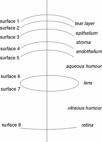
The parameters of the different surfaces are shown in the table below.
|
Surface number
|
Radius of Curvature (mm)
|
Asphericity Q
|
|
Refractive Index
|
Separation
|
|
1
|
7.83
|
-0.26
|
|
|
|
| |
1.333
|
6mm
|
|||
|
2
|
7.46
|
-0.36
|
|
||
| |
1.401
|
50mm
|
|||
|
3
|
7.09
|
-0.46
|
|
||
| |
1.378
|
500mm
|
|||
|
4
|
6.718
|
-0.56
|
|
||
| |
1.357
|
5mm
|
|||
|
5
|
6.34
|
-0.66
|
|
||
| |
1.336
|
3mm
|
|||
|
6
|
11.0
|
-1.0
|
|
||
| |
1.42
|
3.6mm
|
|||
|
7
|
-8.0
|
-0.5
|
|
||
| |
1.336
|
16mm
|
|||
|
8
|
12.0
|
0.0
|
|
||
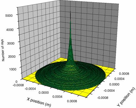
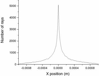
With no scattering in the eye, with this model, the image on the retina is as shown in the figures below.
 |
 |
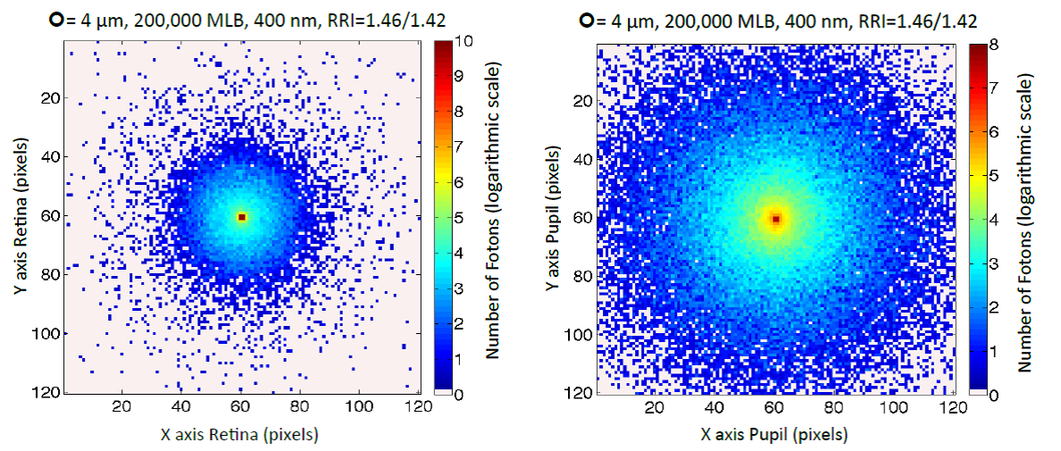
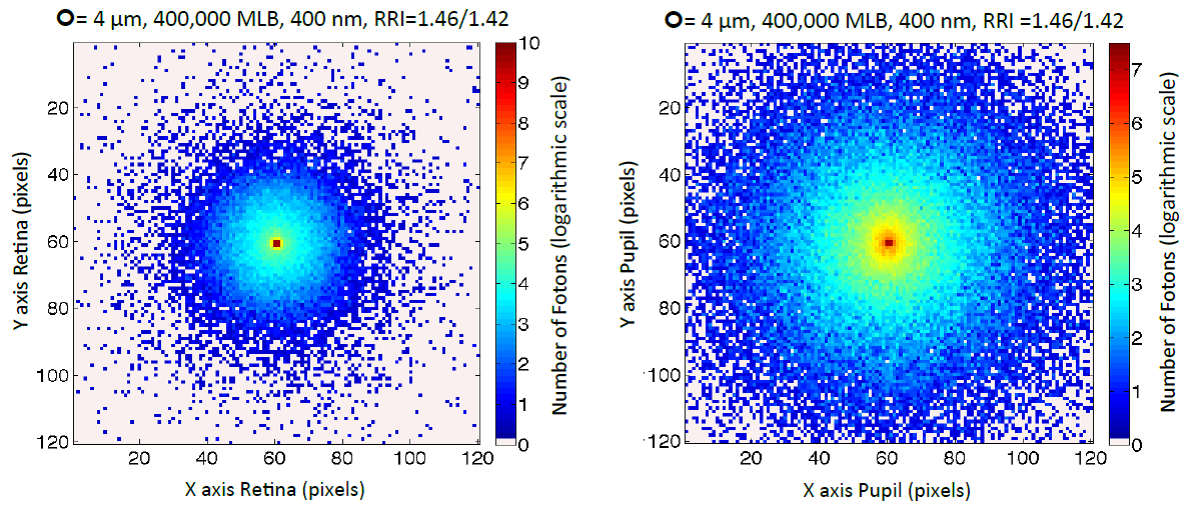
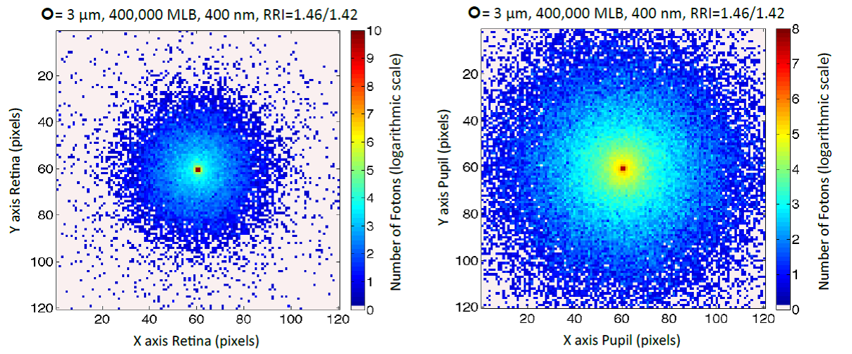
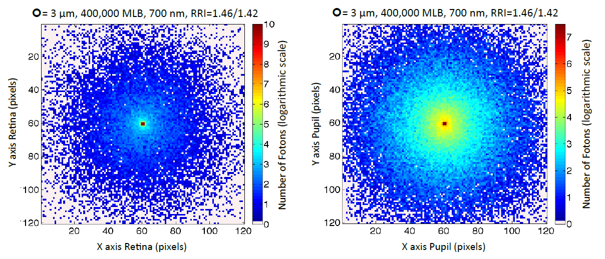
Taking into account the scattering from the cataract particles, we have 3 main variables: cataract particle size, number of cataract particles, and the illuminating wavelength.The first four graphs show scattered light patterns on the retina (left-hand graphs) and the returning photons scattered back out of the eye after travelling through the eye and being backscattered from the retina (right-hand graphs), for 400000 particles in the lens, an illuminating wavelength of 400nm and particles of 2 micron diameter (top) and 4 micron diameter (bottom). The relation of the particle refractive index to the background refractive index is given by the term RRI in the figures.


Publications
1. I. Kelly-Pérez, N.C. Bruce y L.R. Berriel-Valdos, "Modelling image formation on the retina and backscattered light in the human eye with cataracts" International Conference on Applications of Optics and Photonics, Proceedings of SPIE Vol. 8001, 800136, (2011).
2. I. Kelly Pérez, N.C. Bruce y L.R. Berriel-Valdos, "Effect of cataracts on scattering of light in the eye", ICO 2011, Puebla, México, Proceedings SPIE, 8011, (2011), 8011-71
3. I. Kelly-Pérez, N.C. Bruce, L.R. Berriel-Valdos, A. Werner, J.A. Delgado Atencio, "A computational model of the effect of light scattering from cataracts in the human eye", Journal of the Optical Society of America A, 30, (2013), 2585-2594
4. I. Kelly-Pérez, E.M. Mendez-Aguilar, C.G. Treviño-Palacios, N.C. Bruce, L.R. Berriel-Valdos, H. Al-Hohamedi, T. Bende, "Wavelength-dependent scattering in human eyes with cataracts", Journal of Biophotonics, 2018; e201700235, (2018)
| ICAT |